Opticians have various methods to check for macular degeneration. In this article, you will learn how those tests for macular degeneration work. Not every optician uses those tests. Generally available are the following:
- Amsler Grid
- Examination of the fundus of the eye
- Fluorescence angiography
- Optical coherence tomography
Opticians are no medical doctors. This means they can not diagnose eye diseases. But they can check for them and when something appears to be suspicious in the check-up the optician refers to the ophthalmologist and he or she diagnoses, for example, macular degeneration.
Let us start with the simplest way to check for macular degeneration which is the Amsler grid.
Amsler Grid
Amsler grids are grids with a dot in the middle. They can have black lines and a white grid or a black grid and dot and white background. As long as you can not perceive one of the following bullet points the result is good.
- the dot in the center is not visible
- bent or distorted lines
- “holes” or gray veils
- dark or blurred areas
- boxes of different size
The Amsler grid works as follows: If you normally use reading glasses, you must also put them on for the test. Cover one eye and focus on the dot in the center with the other (normal reading distance, 11-15 inches). Then test the other eye, also “one-eyed”, so one eye is always covered and one is tested. Here in the image below you can see how an Amsler grid looks like. It can be presented to you printed out on a sheet of paper or digitally on a screen.
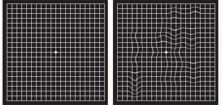
Your optician guides you through the test and focuses your attention on the details that matter to check for macular degeneration. One thing which is very common which is perceived with macular degeneration is the bent lines as you can see on the right side of the illustration above. On the left side, you can see the regular perception of the Amsler grid.
Eximanition of the Fundus of the Eye
During ophthalmoscopy, the optician can look at the back of the eye which is called the fundus. In doing so, he or she looks through a magnifying glass into the interior of the eye under illumination. Here in the image below is an example of what the optician sees.
The dominant brighter spot in the middle of the picture shows the papilla of the eye. There the nerves leave the eyeball. For macular degeneration, the optician looks typically at the little darker spot located to the right to the papilla. Here the visual acuity is the highest. And this is exactly the place where macular degeneration impairs vision.
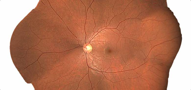
The optician looks for typical structures such as drusen and degenerated, thinned-out tissue that is often visible depending on the macular degeneration. In wet macular degeneration, sprouting vessels, exudates, and hemorrhages are also visible. Usually, the back of the eye is photographed during the ophthalmoscopy to compare the condition with later images. This allows the progression of the disease to be documented.
Optical Coherence Tomography (OCT)
For more precise clarification of macular degeneration, the optician can use optical coherence tomography. With OCT a weak laser is utilized to produce high definition layered images of the retina. This optical coherence tomography is easier to perform than fluorescence angiography because no injection is needed to check for macular degeneration. On top of that, the procedure is very fast and painless for the patient.
There are different methods of how suspicious patterns get detected. Depending on the optician the pictures get evaluated by an opthalmologist, artificial intelligence, or by the optician himself.
Fluorescence Angiography
Fluorescein angiography is a method of visualizing the vessels of the retina and detecting macular degeneration. Due to the needed injection of the fluorescent substance which works as a contrast agent, this method is not for every patient. Especially people which fit the list below should not get fluorescence angiography done:
- renal dysfunction (renal insufficiency)
- Pregnancy
- Breast removal (mastectomy)
- Surgery on the lymph nodes of the armpit
If the contrast agent is injected into a vein in the arm. Shortly after (after 10 to 15 seconds) the contrast agent shows up in the eye and the optician can start with the imaging process. If the eye is now illuminated with short-wave blue light, the dye (contrast agent) lights up. The distribution of the dye in the eye is recorded with the aid of a camera. The evaluation is then done by a medical specialist.
Conclusion
As you can see there are various methods opticians can use to detect macular degeneration. In some cases, the optician can use this procedure on his own while in other cases an ophthalmologist is needed to evaluate the images taken by the optician. By far one of the fastest and cheapest ways your optician can use is the Amsler grid. The test just takes up 2-5 minutes and it tells the optician a lot about your eyes. I personally use this test with every one of my customers.
But it also has its shortcomings. First, the test person needs to interpret the perceived image correctly. With the other ways, the test person is less interaction with the test person is required and a bigger surface of the retina can be checked. When the simple Amsler test screening shows a positive result the optician needs to recommend a further examination by a doctor to see if the test person really has macular degeneration.