Opticians take photos of your eyes for documentation purposes in regards to the inside or outside of your eyes as well as for measurements when new glasses bought. In most cases, those photos are used to measure how the lenses need to be positioned in front of your eyes also known as centration. Here below you, you will find a more detailed list of why opticians take photos and what happens with this data.
- Retinal imaging
- Topography
- Evaluation of contact lens fittings
- Documentation of a progressive change on or in the eye
- Determination of lens opacity
- Centration for glasses
- Documentation of cell defects with contrast agents
Retinal imaging
When opticians take photos of your eyes inner parts retinal imaging is used. Usually, the optician in most countries uses the machine and makes the photos but an ophthalmologist will evaluate the images taken. On the image changes of the retina can be seen or defects that may tell the optician why visual acuity may be reduced.

Retinal imaging is not typically used at optical stores but some use it as a screening tool and if the optician then sees abnormalities at the inner parts of the eyes he will then transfer the customer to an optical doctor. What can the optician see with retinal imaging?
- Damaged blood vessels in your retina can be seen which may cause a loss in visual acuity when not controlled.
- Degenerative changes on your macular can be seen
- The results of intraocular pressure (a damaged optic nerve) can be seen
Topography
In comparison to the paragraphs above the optician solely focuses on the outer part of the eye when a photo is made with a topographer. This device also projects bright circles on your eye. With software tools that analyze the photos, the optician then gets the measurements of the curvature of your eye. This way he is able to tell if glasses or special contact lenses may be a better solution for you.
The topographer is an unbelievably useful tool. It can be used for consultation purposes on which lenses for glasses to buy which contacts might work better or why your vision may be off. Dry eye spots can be seen on your cornea. It is a great tool and I as an optician use this device every day to take photos and to compare them to the measurements we did in the years before in my optical shop.
Evaluation of Contact Lens Fittings
When it comes to evaluating a contact lens fit photos and videos taken of your eyes are also a great way to see how they work. Not only may the optician show you right away what can be improved but he or she can immediately compare the photos from today with the old ones. Documentation is king when it comes to contacts. And a text will never tell you as much as a photo or a video does.
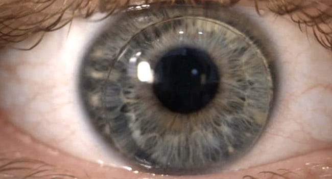
Here in the picture below you can get an idea of what an optician might see.
Documentation of a Progressive Change on or in the Eye
In the paragraphs above I mentioned the documentation which is very important. Herewith the eye shown on the image below the customer wants to know if the condition (in this case pterygium) got worse. The optician can see immediately by comparing the images if the condition changed.
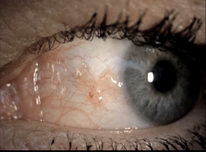
And usually things change when it comes to the eyes. usually over the years the recommendations change in regards to the contacts fitted because of the lower quality tear film. Oftentimes the optician can see with an simple image if the opacity of the eyes lens is still good or if the customer should see a doctor because new glasses will not do the job.
Determination of Lens Opacity
The optician can check the opacity of the lenses. Different technology can be used to check the opacity. One example is the slit lamp where a tiny light beam shine through the parts of your eye. When the light passes the different structures the optician can tell if the lens already produces a blurry image and rather should be replaced.
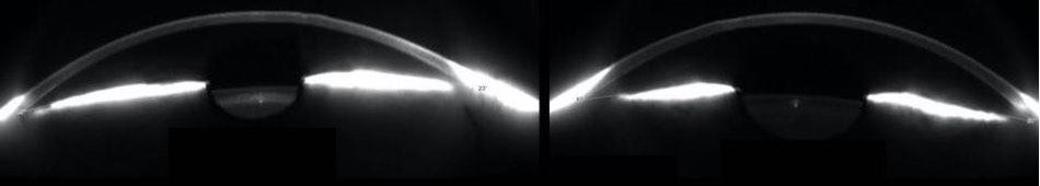
The second and more advanced way to check the opacity of the lens is a scheimplug camera which scans the anterior part of your eyes. With this technology, the optician gets up to 25 000 data points to calculate topographic corneal thickness, corneal curvature, anterior chamber angle, volume, and height. This machine is incredible and it makes my day every time I use this device.
Here in the picture above you can see the differences of the lenses in the middle of the picture. The left one looks a little opaque while the image on the right side shows a totally transparent eye lens.
It is especially useful to take images with it when it comes to very complicated contact lens fittings and highly specialized lenses need to be fitted.
Centration for Glasses
The centration is oftentimes also done with a photo. Here the preadjusted frame is documented with your face. Afterward, the optician uses this photo with software to set reference points in order to measure where he needs to position the optical center of the lenses in your new glasses.
Depending on the lenses used a photo from the front in combination with a photo from the side will be used. This way the optician also can measure the vertex distance and how much the glasses are tilted in the face.
Documentation of Cell Defects With Contrast Agents
Contrast agents are an important part of evaluating the eye’s surface. Therefore different contrast agents like fluorescein, lissamine green, or Bengal rose can be used to check the surface of a customer’s eye for defects. Those defects can come from too much bearing of the contact lens or a foreign body that scratched the eye.
Different contrast agents show defects on different parts of the eye. I have another article here for you about why exactly an optician might put dye in your eyes.
This is basically why opticians take photos of your eyes. It is all about measurements and documentation around the eye and the glasses or contacts. After writing this opticians really take a lot of photos during the day. I hope you found the information you were looking for about why opticians take so many photos.
I wish you a great day.