Conventional cataract surgery is the most common surgical procedure in the world and has been performed according to the same principle for about 40 years.
During cataract surgery, the clouded, natural lens of the eye is removed and replaced with an artificial intraocular lens (= lens inside the eye). This surgical procedure is extremely routine and standardized.
In most cases, the surgery is performed on an outpatient basis. Only in special cases is the patient treated as an inpatient.
Even if both eyes are affected by cataract, as a rule only one eye is operated on at a time – the more severely affected one first.
Of course, cataract surgery has been supplemented with current technologies and methods. Currently, the most modern and gentle procedure is as follows.
Cataract surgery Explained in 6 steps
Anesthesia
Before the operation, the eye is anesthetized by means of drops (local anesthesia). Many patients receive an additional sleep anesthesia (sedation) to calm them down.
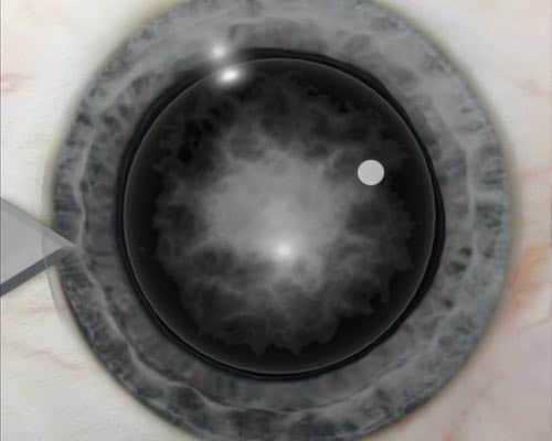
Creating Surgical Access
The operation is minimally invasive. Whereas in the past incisions of approx. 11-13mm were required as surgical access in the sclera, today only an approx. 2-3mm corneal incision is required. The eye surgeon makes this incision with a fine scalpel.
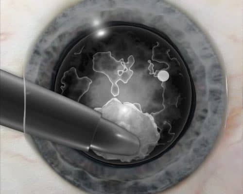
Phacoemulsification
Now the phacoemulsification is performed: the anterior lens capsule is incised and opened circularly (capsulorhexis). A hollow needle is inserted into the anterior chamber of the eye.
Using pulsed ultrasound shock waves and surgical instruments, the hardened lens nucleus is crushed and aspirated through the hollow needle along with the softer lens cortex.
Depending on the anatomical characteristics, as much of the capsular bag as possible is left in the eye to serve as a fixation for the intraocular lens.
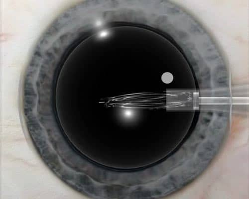
Implantation of the Intraocular Lens
The artificial intraocular lens is then implanted into the eye. Since it is much larger than the surgical access, it is inserted into the eye folded with an injector and only unfolded and aligned in the eye. The intraocular lens has elastic stirrups with which it fixes itself in the remaining part of the capsular bag after alignment.
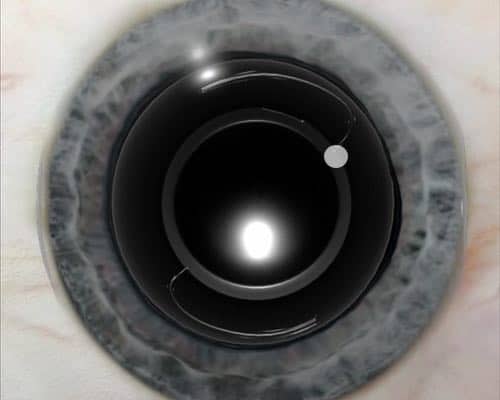
Alignment of the Intraocular Lens
If a toric intraocular lens is implanted, the most accurate axial alignment possible is important for postoperative vision. Therefore, the optimal alignment angle is marked on the patient’s eye before surgery and the lens is aligned accordingly.
The positioning of the lens is therefore in many cases not only very sensitive in terms of tilt, but also the rotation of the lens must be taken into account.
Completion of the Operation
Due to the minimally invasive procedure, the small incision in the eye does not require stitches and heals independently.
The entire cataract surgery takes only about 15 minutes. During the surgery the patient does not feel any pain.
After Cataract Surgery
The patient receives an ointment dressing to protect the operated eye from external influences. For monitoring purposes, the patient remains in the eye clinic for a few hours and then – if no complications occur – is allowed to go home.
The day after surgery, the bandage can be removed during the day. Even though the eye still needs some time to get used to the new lens, many patients are already happy about their regained vision at this point. The ointment bandage should be worn at night for about another week. In the following weeks, regular check-ups by the attending ophthalmologist are necessary.
Patients often report a foreign body sensation or slight scratching of the eye that lasts for a few hours or a few days. This is normal.
After 5-7 days, most patients can either resume reading or watching television without glasses. Fitting of new glasses can usually be done after about 4-6 weeks.
In 95% of cases there are no complications with cataract surgery. If you would like to know more about the complications, we have an article for you here.
As you could see, cataract surgery is a routine procedure nowadays. Complications are rare. However, to avoid complications, you should carefully discuss the differences in cataract surgery. For example, you can choose whether you want to have sharp near vision without glasses, but blurred far vision or vice versa.
With single vision intraocular lenses you can usually see very sharply but only at one distance. Another option is multifocal intraocular lenses, for example, which may only give you 90% sharpness instead of full vision. However, you might then be able to do without distance and reading glasses in most situations.
As you can see, there are many possibilities. So prepare for the appointment and think about what distances you would like to see sharply in everyday life without glasses after cataract surgery.